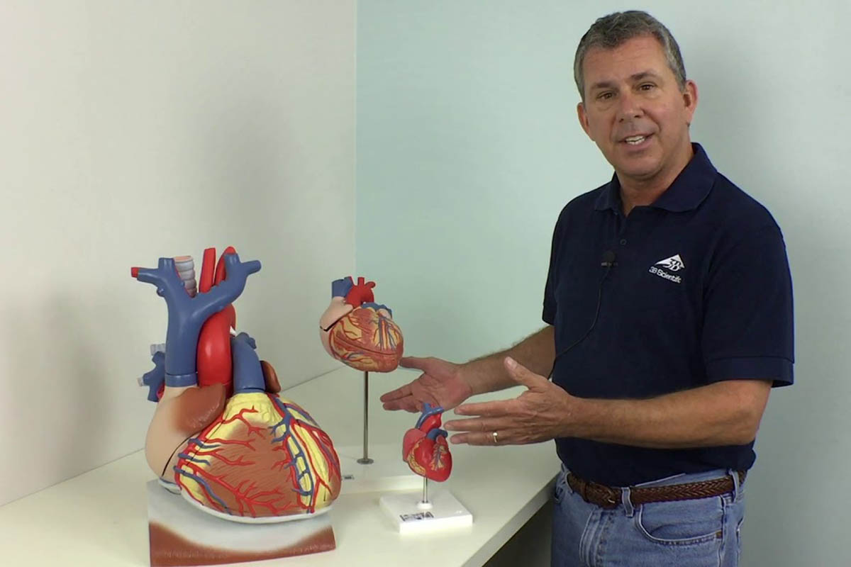
Physical models of internal organs are useful in clinical practice for a variety of reasons, including planning for difficult surgeries. To replicate the complexities of unique patient anatomies, the heart is typically modeled using 3D printing.
A team from Carnegie Mellon University has successfully modeled a patient’s heart through 3D printing. The team used alginate, a seaweed-derived material that closely resembles the texture of cardiac tissue. This implies that cardiac surgeons may practice their procedures on the model, slicing and suturing it in anticipation of real-life surgery.
The team’s leader, Adam Feinberg, said that they are now able to build a model that they can use to do conceptual preparation and physical practice. The surgeon can stimulate it and make it react like real tissue. This will give them practical experience in that environment before they get into the surgery room.
The model was created using a technique known as the FRESH technique, which stands for Freeform Reversible Embedding of Suspended Hydrogels. It works by introducing bioinks into a soft hydrogel before it is heated to expose the final form of the object.
The technique was improved and extended in this latest advancement to create complete 1:1 replicas of real patient hearts relying on tomography data collected from MRI scans.