BIOMONITOR IIIm

Biotronik has declared clearance and approval by the FDA of an important Data Sensor in the latest Biomonitor IIIm injectable cardiac monitor (ICM). The sensor detects body temperature changes that may also be associated with fever.
The connection between vital signs, such as body temperature and cardiac arrhythmia, and health problems has been long known. Temperature increases, in particular, can cause a rise in heart rates and, as a result, a reduction in the patient’s activity level.
With the Data Sensor, doctors can access comprehensive updates on critical data to assist them in remotely tracking patient’s overall …

Enhancing Structural Heart Disease Treatment

CardioCare is presented as a new intelligence-based approach that aims to assist clinicians in identifying and treating patients with chronic heart disease. It is already used in more than 50 clinics and hospitals across the country.
It’s not uncommon for structural heart disease to go undiagnosed or undertreated. The secret to successfully treating structural heart disease is an accurate diagnosis of the disease and the problematic area within the heart. There is also a need for thorough follow-up.
CardioCare was established to assist physicians in the diagnosis and treatment of such patients. For example, the system …

Using Synthetic Antibody to Limit COVID-19 Spread

Governments and companies have started distributing a few varieties of COVID-19 vaccines around the globe. Even so, researchers at the University of California (UCSF) have formulated an innovative method that may provide a somewhat more effective way to curb the development of coronavirus inside the human body.
The researchers developed an artificial antibody that keeps the COVID-19 virus from sticking to human cells. These artificial antibodies are based on the microscopic antibodies that are produced by animals like llamas and camels, to defend themselves from coronavirus varieties.
The antibodies attack the spikes found on…

Ultra Precision 3D Human Heart Models

Physical models of internal organs are useful in clinical practice for a variety of reasons, including planning for difficult surgeries. To replicate the complexities of unique patient anatomies, the heart is typically modeled using 3D printing.
A team from Carnegie Mellon University has successfully modeled a patient’s heart through 3D printing. The team used alginate, a seaweed-derived material that closely resembles the texture of cardiac tissue. This implies that cardiac surgeons may practice their procedures on the model, slicing and suturing it in anticipation of real-life surgery.
The team’s leader, Adam Feinberg, …

Implantable LVAD for Heart Failure

Due to the serious damage to the heart muscle, persons with chronic heart failure frequently require a cardiac transplant. As these patients wait for a transplant, LVADs or Left Ventricular Assist Devices are often used to support the heart in pumping blood across the body.
Also, these devices are widely used in the short term to aid the hearts of patients who have undergone heart surgery. Besides, they are a long-term alternative for patients with heart failure but are unable to receive a transplant.
According to Greg Arber from Corvion, there is still a greater need for more LVADs. From around 100,000 to 300,000 people who …

Powerful MRI from GE Healthcare Cleared by FDA

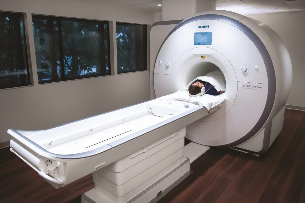
The FDA has cleared the Signa 7.0T MRI scanner from GE Healthcare, which is currently the most powerful tool available for clinical purposes. While the magnetic field of a traditional high-end scanner is 3 Tesla, the magnetic field of this latest device goes all the way up to 7 Tesla. Consequently, it provides an unparalleled view of extremely detailed anatomy.
Although the SIGNA 7.0T is optimized for neurological tasks, it can also be useful for detailed imaging of joints, metabolism, and movements that would be difficult to see with a less powerful device. Aside from clinical uses, the scanner may be used for research purposes as …

Researches Capture Brain Spheroid’s Internal Structure with Digital Imaging

The Wyss Center and associates researchers have come up with novel imagery and labeling techniques for displaying the brain spheroids’ internal structure. They have also been able to observe the anatomy of single neurons in three dimensions (3D).
Brain spheroids, also known as “Mini brains  by researchers,” are a set of various types of brain cells derived from activated pluripotent stem cells. According to researchers, brain spheroids can be used for a variety of purposes, including drug development, drug testing, and neurological disease studies.
For researchers to examine their structural properties, they must …

FDA Approves Double Black Imaging’s Display for Breast Imaging

Following FDA approval, Double Black Imaging, a Plymouth-based firm that focuses on medical screens and displays, has released an outstanding 12-megapixel monitor designed for use in digital mammography.
The Gemini 12 MP features a screen size of 31 inches. Unlike the previously used two 5-megapixel displays, it can capture 2 different tomography or mammography exams, making it easy to compare them on just one screen. This eliminates the intrusive plastic bezel in between, making it convenient for the doctor.
The monitor includes a high-speed graphics controller that is designed for processing tomo image. It also has custom-…

Strados Labs’s RESP Lung Sound Monitoring Device Received FDA Approval

Strados Labs, a Philadelphia-based medical technology firm, has received FDA approval to bring its RESP lung sound monitoring device to hospitals, clinics, and doctors. The RESP device has been used in clinical studies to determine how clinicians can use it to monitor patient reports, therapies, and other treatments that relate to lung sound alterations.
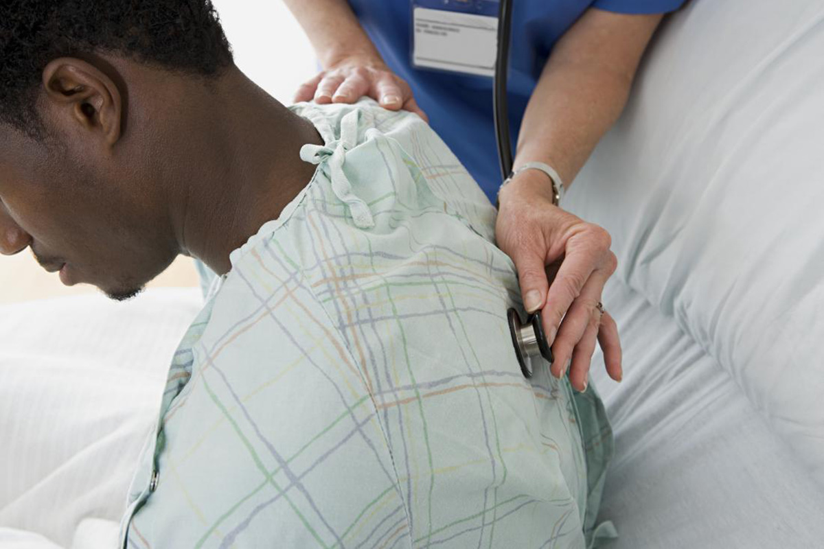
The device works by placing it on the chest of the patient. It then notes any typical lung sounds like coughs just like a stethoscope do. The results are then recorded and stored digitally in a HIPAA compliant way so that clinicians can easily access the data for monitoring and …

Detecting COVID-19 with Microfluidic Mobile Phone Extensions

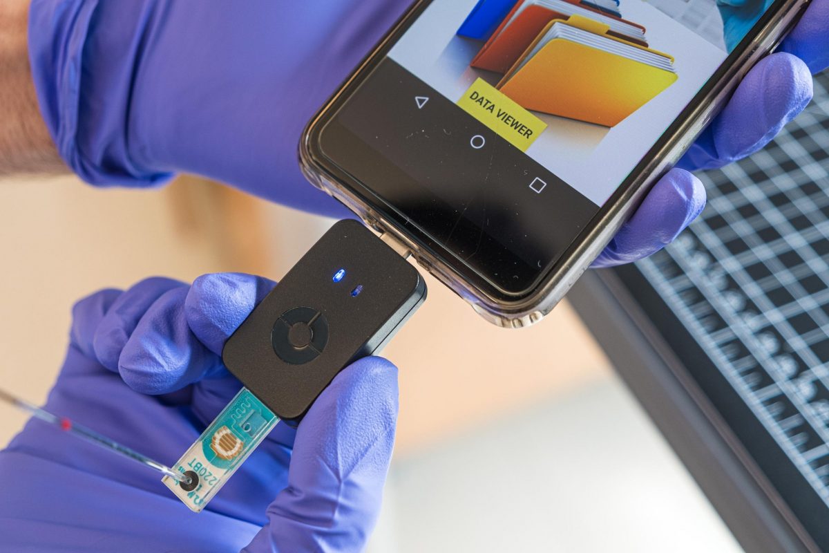
The battle against COVID-19 remains, and monitoring is one of our most convenient and practical tools for halting the virus’s transmission. In this case, Rice University researchers have created a tool that uses the microfluidic method to detect COVID-19 in a blood sample and show the results once inserted into a mobile phone.
The test uses antibody-studded magnetic nanobeads to recognize COVID-19’s nucleocapsid protein. It takes less than an hour to get results for the test. The device was able to detect very low concentrations of nucleocapsid protein during clinical trials, indicating that it could be used as a …