The Wyss Center and associates researchers have come up with novel imagery and labeling techniques for displaying the brain spheroids’ internal structure. They have also been able to observe the anatomy of single neurons in three dimensions (3D).
Brain spheroids, also known as “Mini brains by researchers,” are a set of various types of brain cells derived from activated pluripotent stem cells. According to researchers, brain spheroids can be used for a variety of purposes, including drug development, drug testing, and neurological disease studies.
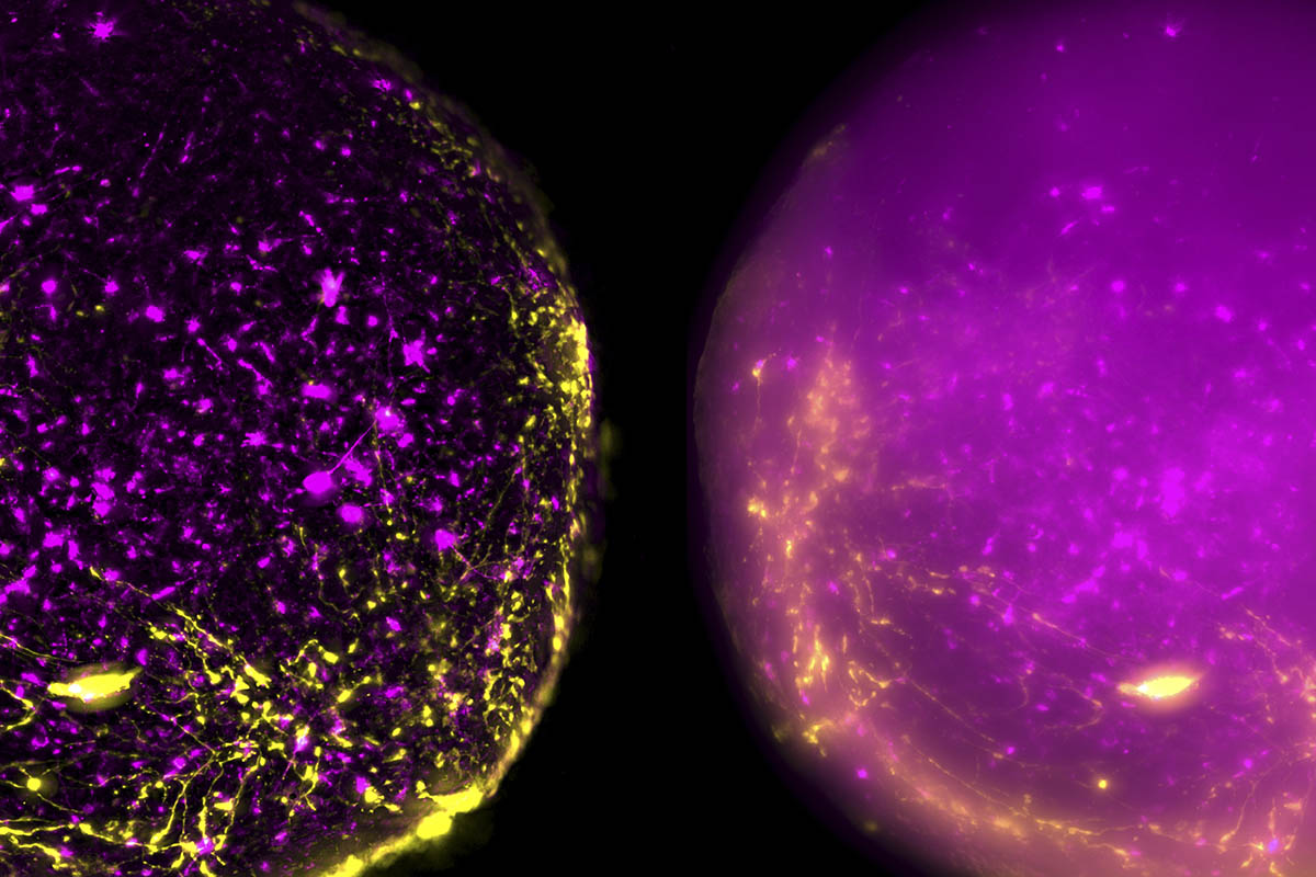
For researchers to examine their structural properties, they must cut them into small parts and position them on microscope slides. However, the spheroids would be more beneficial for researchers as a learning tool, including in drug production if their structure can be reliably measured without cutting them into fragments for microscopy.
Another scientist involved in the process, Dr. Subashika Govindan, explained that to examine the inside of a spheroid, they need to slice it thinly and view it on a slide under a microscope.
According to Govindan, this is a time-consuming procedure that can harm the sample. Researchers have now generated high-resolution 3D image data of single neurons inside intact spheroids for the first time, exposing their extraordinary intricacy.